Medical Exhibits - Demonstrative Evidence Expert Blog - MediVisuals

Intra-articular Fractures Explained

Posted by Delia Dykes on Wed, Oct 5, 2011

By: Robert Shepherd MS, Certified Medical Illustrator, Vice President and Director of Eastern Region Operations, MediVisuals Incorporated

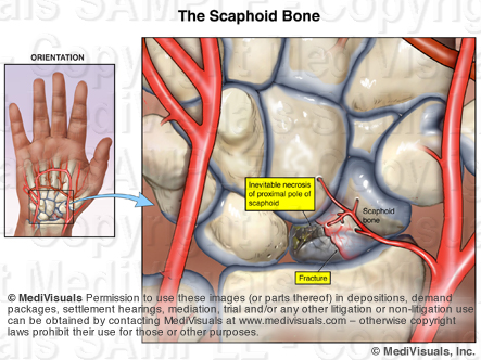
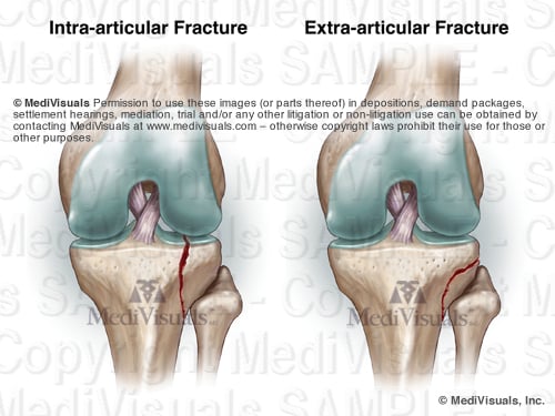
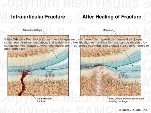
Intra-articular fractures are simply fractures that involve a joint space (see below figure). While intra-articular fractures appear very similar to those that do not involve a joint space (extra-articular fractures), intra-articular fractures are significantly more serious because they are associated with a much greater incidence of long-term complications.


Image1 TwoFracsSTAMPED


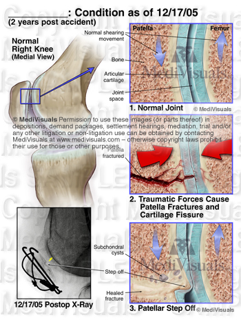
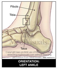
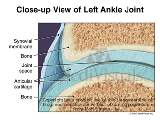
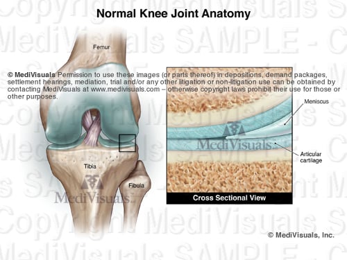
In order to appreciate why intra-articular fractures can be so problematic, a fundamental understanding of a typical joint is helpful. The following images show a knee joint. With the exception of a meniscus, almost all moveable joints are similar to the knee joint in that the joints are lined with a thick, shock-absorbing articular cartilage adherent to smooth, bony surfaces that allow pain-free movement.


Image2 NormJointSTAMPED

When a fracture involves the articular surface of one or more bones of a joint, the articular cartilage and smooth articular surface of the bone are disrupted. In order for joints to have the best chance of proper joint function after healing, physicians go to greater effort to make sure the bony surfaces are properly aligned and that the joint is properly immobilized than they would with a similar fracture that is extra-articular. Even with the best fracture alignment and joint immobilization, subtle disturbances in the joint surface and the natural bone reformation that take place during healing can result in uneven joint surfaces and injury to the overlying articular cartilage (see the below illustration). Because of the abnormalities of the injured and healed joint surface, natural movement of the joint can also damage the articular cartilage of the opposing joint surface. Over the course of time, these injuries self-perpetuate and may necessitate arthroscopic debridementchondroplasty or even joint replacement.


Image3 Frac AfterHealSTAMP

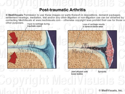
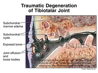
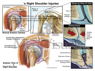
It is also important to realize that a fracture needs not enter a joint to result in injury to the articular surfaces and begin the self-perpetuating post-traumatic breakdown of the joint surfaces (post-traumatic arthritis). As shown in the below illustrations, joint trauma without a diagnosable fracture of any type can injure the smooth, shock-absorbing articular cartilage, with or without microfractures of the underlying bone. This can result in partial or total loss of the articular cartilage and in uneven "bone-on-bone" articulation that severely decrease range of motion and result in debilitating joint pain.


Image4 ArthritisSTAMPED2

Topics: knee replacement, arthritis, joint, ankle, medical exhibits, medical-legal-illustration, MediVisuals, medical exhibit, facet joint, personal injury

Complications Associated with Fracture Injuries

Posted by Trisha Haszel Kreibich on Wed, Jun 8, 2011

Several long term or permanent complications may result from a fracture injury. Traumatic arthritis may be one of the most common long term debilitating complications. (See the previously posted article "Breaking Down Traumatic Arthritis" for more information.) These complications may even require additional surgical procedures.

Normally, fractures begin healing by forming a callus which is then replaced by bone. With a nonunion (see image below), the callus is replaced by fibrous tissue instead of by bone. This is a painful condition that is most frequently treated by surgery to remove the fibrous tissue, debride the surrounding bone and re-approximate the remaining fragments (sometimes with bone graft material and fixation devices).

Nonunion fracture

Malunion is another frequently occurring complication of fractures. With malunion, the bones heal, but they do so at an unusual angle that can effect range of motion and/or cause pain (see image below). Treatment typically involves refracturing or cutting the bones, realignment, and fixating them with hardware so that they heal in the correct position.

Alignment Misalignment of Wrist
Intra-articular “step-offs” are a complication of intra-articular fractures. A “step-off” is a type of malunion in which the bone heals, but it heals so that the joint surfaces are not aligned in a smooth continuous surface. The illustration below shows a fracture through the patella that healed with a painful “step-off.”

Patella Fracture Knee Joint

Like all organ systems, bones require a blood supply. If the blood supply is disrupted, the bone can die and collapse. This condition is known as avascular necrosis. The scaphoid is a bone in the wrist that is frequently affected by avascular necrosis. In the illustration below, a fracture is seen through the scaphoid and its blood supply.  As a result, the small fractured fragment loses its blood supply and over time, becomes necrotic. Treatment would likely involve fusion of the wrist bones. Other bones that are particularly vulnerable to avascular necrosis include the femoral head (hip joint) and the talus (ankle joint).

Scaphoid Avascular Necrosis

Ossification of the surrounding soft tissues is another complication of fractures. This condition is referred to as “heterotopic ossification” and/or “myositis ossificans”. With this condition, the bone cells that form to heal the fracture extend out into the surrounding tissues (see image below) resulting in pain and decreased range of motion. Treatment involves surgery to excise the excessive bone growth.

Heterotopic Ossification Thigh Fracture

Topics: malunion, step-off, arthritis, ankle, fracture, nonunion, avascular necrosis, ossification, medical exhibit

Breaking Down Traumatic Arthritis

Posted by Trisha Haszel Kreibich on Tue, Dec 15, 2009

By: Robert Shepherd MS, Certified Medical Illustrator, Vice President and Director of Eastern Region Operations, MediVisuals Incorporated

Fractures can result in several long term or permanent complications that can necessitate additional surgical procedures. One of the most common long term debilitating complications is traumatic arthritis. Traumatic arthritis can affect almost any moveable joint in the body. To explain traumatic arthritis more in depth, we will be focusing on the tibiotalar (ankle) joint, as shown in the illustration to the right.

arthritis-tibiotalar-joint

As like most moveable joints, the tibiotalar joint consists of smooth articular bone covered by thick, shock-absorbing articular cartilage.

arthritis-joint-normal

During trauma, the joint surfaces can be driven together resulting in injuries to the cartilage and microfractures of the articular surface (even without obvious intrarticular fracture).

arthritis-injury-joint

The bone and cartilage then undergo changes that result in the progressive breakdown of the joint. As the process advances, the joint becomes painful. In most cases, the only treatment options are joint replacement or fusion.

arthritis-degeneration-joint

The acromioclavicular (AC) joint also frequently falls victim to traumatic arthritis. As the AC joint enlarges (hypertrophy) it impinges on the rotator cuff, which is referred to as subacromial impingement. This can cause irritation or tearing of the rotator cuff and is most often treated by AC joint resection and subacromial decompression.

arthritis-AC-joint-shoulder


ZSN7H32E27UR

Topics: arthritis, joint, ankle, medical-legal-illustration, MediVisuals