Medical Exhibits - Demonstrative Evidence Expert Blog - MediVisuals

Medical Legal Illustration and Animation - Cross Sections

Posted by Tara Rose on Mon, Jun 18, 2012

By: Robert Shepherd MS, Certified Medical Illustrator, Vice President and Director of Eastern Region Operations,MediVisuals Incorporated

Long before science had advanced to allow imaging of the body in sectional views by computed tomography (CT) and magnetic resonance imaging (MRI), medical illustrators were illustrating the body in sectional views because these views are the best way to appreciate some anatomical relationships. 

Medical illustrators, physicians, and others who have studied anatomy are familiar with sectional views of the body and appreciate the value of these views in explaining the relationship of anatomical structures. However, accomplished and well respected jury consultants and non-medical illustrator legal graphics experts have expressed concerns that sectional views may be difficult for some jury members to understand. These individuals' opinions are valuable to those of us in the legal graphics business, and I agree with their opinions that, when other views can communicate a particular relationship message equally as well or better, sectional views should be avoided. I also believe most of these experts will agree that there are times and places in which sectional views of anatomy are the best way to appreciate some anatomical relationships. Granted, there have been times when we have been working on specific cases and experts have insisted that sectional views be absolutely and unconditionally avoided. Unfortunately, in these situations the experts were unable to suggest a more effective view to communicate the relevant anatomical relationships (at least in a way that was practical in terms of time and expense). That being the case, sometimes the sectional views were used despite the input of the experts, and at other times, the relationships of the structures had to be explained without the benefits of graphics.

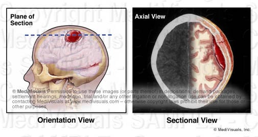
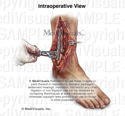
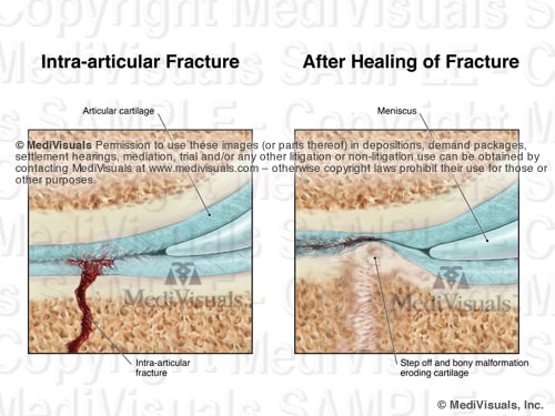
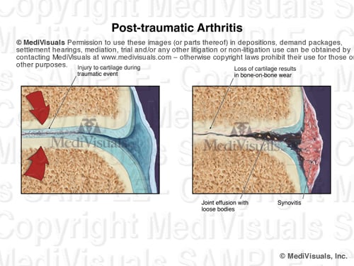
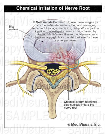
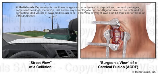
A way to perhaps explain how sectional views help decision makers appreciate relevant anatomical and pathological relationships is to compare them to aerial views or photographs of the scene of a collision.  Space is defined in three planes. Only two of these planes can effectively be demonstrated in a two-dimensional rendering. For example, aerial views have long been used to help explain the positions of vehicles and structures that simply can't be appreciate from "street views". When viewing the scene of a collision from a "street view", one can appreciate vertical and horizontal distances, but not depth; distances close to and far from the viewer's perspective are very difficult to appreciate (see the below figures). By comparison, when viewing an operative site through a "surgeon's view", vertical and horizontal distances can be appreciated, but the depth of the incision and the relationships of the various structures within and around the incision are very difficult or impossible to appreciate.


Cross Section Blog image1 REVISED


The "aerial view" of the collision scene allows the viewer to appreciate distances in two geographical planes as well (distances right and left, and toward and away from the "street view," but the ability to appreciate up and down is lost). Also, the locations of relevant structures or vehicles that may have been obstructed by nearby structures (such as buildings trees, signs, or other vehicles) can now be appreciated. Similarly, a sectional view of anatomy can help decision makers appreciate depth relationships of structures. Or, a sectional view of a step in a surgical illustration can allow the viewer to appreciate the depth of the surgery as well as the additional structures that may have been injured (or at risk of injury) during the invasive procedure.  These specific depth  relationships could not be appreciated from the "surgeon's view" of the same surgery shown in the above illustration.


Cross Section Blog image2 REVISED


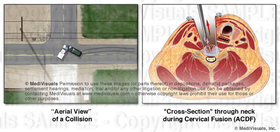
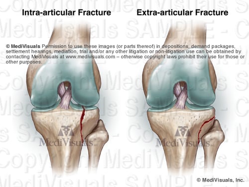
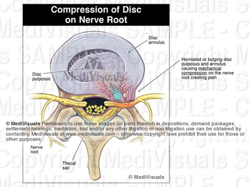
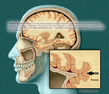
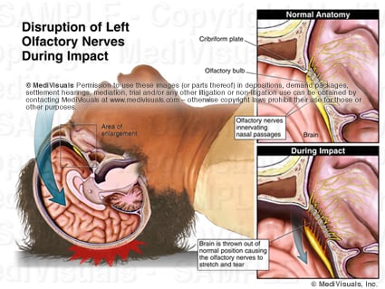
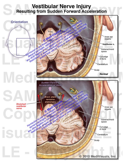
Exhibits developed to help explain the invasive nature of a surgery and the disruption of the soft tissues during operative procedures are critical. For that reason, sectional views are critical in aiding a testifying physician to explain these issues. For example, the exhibit panel that demonstrates an anterior cervical discectomy and fusion (ACDF) that does not include a cross-section through the neck fails to emphasize the depth of the incision and disruption of tissues (essentially all the way to the center of the neck). This depth simply cannot be appreciated in a "surgeon's view".

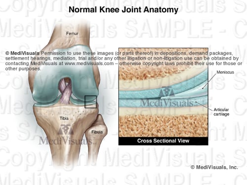
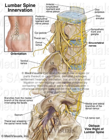
In order to appreciate cross-sections, orientation views that show the level and direction of the section are helpful (see below), or when time, budget, and presentation format (digital as opposed to a physical panel) allow, a short animation showing the sectional view actually coming out of the orientation view such as MediVisuals' "Scan SelectorTM" can be used.


cross-section plane of brain hematoma

Topics: intervertebral disc, coup-contracoup, medical-illustrator, trial exhibit, disc herniation, degenerated disc, disc bulge, trauma, hematoma, traumatic-brain-injury, medical exhibits, medical-legal-illustration, disc injury, brain, TBI, medical expert, intracranial, surgery, MediVisuals, medical exhibit, personal injury, spinal injury

Disc-Osteophyte Complex Explained

Posted by Delia Dykes on Wed, May 2, 2012

By: Robert Shepherd MS, Certified Medical Illustrator, Vice President and Director of Eastern Region Operations, MediVisuals Incorporated

Individuals who develop new or suddenly worsening symptoms consistent with nerve root or spinal cord impingement following a traumatic event are sometimes diagnosed with “disc-osteophyte complexes”. The term “disc-osteophyte complex” generally refers to abnormal extension of intervertebral disc material that accompanies immediately adjacent osteophyte formation at the vertebral body margin (see the below figure). It is important to note (as shown in the illustrations) that the disc almost always extends further than the osteophytes into the neural foramen or spinal canal to irritate or impinge upon nerve roots or the spinal cord.


DiscOstCompBlogASTAMP


Occasionally, individuals who are evaluated shortly after a traumatic event are found to have disc-osteophyte complexes. Because a minimum of several weeks is required for osteophytes to form as a result of a traumatic event, defendant insurance companies may argue that the presence of osteophytes so soon after the traumatic event in question may prove that the plaintiff’s injuries preexisted the traumatic event. Since it is the disc pathology extending beyond the osteophytes that is the actual cause of the nerve root or spinal cord irritation and inflammation, the defense’s arguments are not valid. As shown in the illustrations below, the sequence of events that typically takes place in these cases is that the plaintiff had minimally symptomatic or asymptomatic disc osteophytes prior to the traumatic event in question. During the traumatic event, the disc sustains trauma that results in worsening of the disc pathology while the osteophyte portion of the osteophyte/disc complex remains essentially unchanged. This worsening of the disc pathology in turn results in new or increased irritation or impingement of the neural elements.


DiscOstCompBlogBSTAMP

Topics: intervertebral disc, medical-illustrator, trial exhibit, disc herniation, disc bulge, trauma, medical exhibits, medical-legal-illustration, disc injury, MediVisuals, medical exhibit, personal injury, spinal injury, osteophyte, cervical strain

Intra-operative Trauma: The Overlooked Injuries

Posted by Delia Dykes on Wed, Oct 19, 2011

By: Robert Shepherd MS, Certified Medical Illustrator, Vice President and Director of Eastern Region Operations, MediVisuals Incorporated

The surgical trauma that a plaintiff has to undergo after the initial bodily injuries following a traumatic event are always major points of emphasis when arguing damages in a personal injury case.  This is certainly the situation with cases that involve broken bones that require invasive surgical procedures to realign broken bone fragments ("reduce") and secure ("fixate") the bones with hardware to keep them properly aligned during healing.  Too often, however, the emphasis is solely on the effects on the bones from these "Open Reduction and Internal Fixation" (ORIF) procedures, and very little emphasis is placed on the surgical disruption of the soft tissues that takes place during these procedures.

In a case involving ORIF of a distal fibula (a.k.a. lateral malleolus) fracture, in order to emphasize the surgical trauma endured by a plaintiff, an attorney may have a visual prepared of a postoperative X-ray.  The visual may consist of only a postoperative X-ray or a print of the X-ray with a corresponding illustration (see the below figure). 


SurgTraumBlog01STAMPED

The above images are certainly helpful, but fail to address the intra-operative trauma to the soft tissues that is required to gain access to the bone fragments.  For that purpose, intra-operative illustrations that truthfully depict the soft tissue disruption should be considered (see the below figure) or even an animation showing the procedures such as the one at this link: http://www.medivisuals.com/fibularplatingORIF.aspx

Illustrations or animations that at least touch on the soft tissue disruption allow testifying physicians the opportunity to explain the many tissues traumatized during the procedure and allow insurance adjustors, mediators, and jurors an opportunity to take these additional injuries into consideration when determining the severity of a plaintiff's entire injuries.


SurgTraumBlog02STAMPED


Many attorneys considering realistic illustrations such as the one above, express a concern that judges may not allow the images to be used because they are too "graphic" or "inflammatory".  Certainly, counsel should make themselves aware and consider the preferences of certain jurisdictions and specific judges before determining whether an illustration should be developed that realistically depicts injuries or whether diagrammatic (cartoon-like) illustrations should be developed instead.  There are a number of very good arguments to support the use of "realistic" illustrations over "cartoons".  Those arguments as well as other discussions regarding illustration styles will be addressed in future blogs.


Topics: demonstrative evidence, soft tissue injury, trial exhibit, trauma, ankle, medical-legal-illustration, surgery, fracture, MediVisuals, medical exhibit, personal injury

Intra-articular Fractures Explained

Posted by Delia Dykes on Wed, Oct 5, 2011

By: Robert Shepherd MS, Certified Medical Illustrator, Vice President and Director of Eastern Region Operations, MediVisuals Incorporated

Intra-articular fractures are simply fractures that involve a joint space (see below figure). While intra-articular fractures appear very similar to those that do not involve a joint space (extra-articular fractures), intra-articular fractures are significantly more serious because they are associated with a much greater incidence of long-term complications.


Image1 TwoFracsSTAMPED


In order to appreciate why intra-articular fractures can be so problematic, a fundamental understanding of a typical joint is helpful. The following images show a knee joint. With the exception of a meniscus, almost all moveable joints are similar to the knee joint in that the joints are lined with a thick, shock-absorbing articular cartilage adherent to smooth, bony surfaces that allow pain-free movement.


Image2 NormJointSTAMPED

When a fracture involves the articular surface of one or more bones of a joint, the articular cartilage and smooth articular surface of the bone are disrupted. In order for joints to have the best chance of proper joint function after healing, physicians go to greater effort to make sure the bony surfaces are properly aligned and that the joint is properly immobilized than they would with a similar fracture that is extra-articular. Even with the best fracture alignment and joint immobilization, subtle disturbances in the joint surface and the natural bone reformation that take place during healing can result in uneven joint surfaces and injury to the overlying articular cartilage (see the below illustration). Because of the abnormalities of the injured and healed joint surface, natural movement of the joint can also damage the articular cartilage of the opposing joint surface. Over the course of time, these injuries self-perpetuate and may necessitate arthroscopic debridementchondroplasty or even joint replacement.


Image3 Frac AfterHealSTAMP

It is also important to realize that a fracture needs not enter a joint to result in injury to the articular surfaces and begin the self-perpetuating post-traumatic breakdown of the joint surfaces (post-traumatic arthritis). As shown in the below illustrations, joint trauma without a diagnosable fracture of any type can injure the smooth, shock-absorbing articular cartilage, with or without microfractures of the underlying bone. This can result in partial or total loss of the articular cartilage and in uneven "bone-on-bone" articulation that severely decrease range of motion and result in debilitating joint pain.


Image4 ArthritisSTAMPED2

Topics: knee replacement, arthritis, joint, ankle, medical exhibits, medical-legal-illustration, MediVisuals, medical exhibit, facet joint, personal injury

Association of Medical Illustrators Recognizes the Best in Medical-Legal Illustration

Posted by Delia Dykes on Wed, Aug 10, 2011

By: Delia Dykes, MS

Each year, the Association of Medical Illustrators has a juried Salon at its national conference (this year in Baltimore, at the end of July). Unlike most "art" competitions, the judging criteria are not just based upon the aesthetics of the illustration -- instead, the illustrations are most heavily judged by how well the illustrations communicate a message and upon the illustrations' anatomical and medical accuracy.  MediVisuals was honored once again this year to receive the Award of Excellence and the Award of Merit in Medical Legal illustration.

The below illustration, created primarily by Paul Gross, MS, received the Award of Excellence for effectively demonstrating a surgical procedure to repair severe facial lacerations resulting from a dog bite.  The illustrations were created for Susan M. Bourque of Parker & Scheer, LLC in Boston. The exhibit assisted Parker & Scheer in obtaining a substantial recovery on behalf of the client.

blog awards paul

The Association's Award of Merit was bestowed to Cynthia Yoon, MS.BMC, the primary illustrator for a series of three exhibits demonstrating severe facial injuries and surgical repairs resulting from an awning pole from a passing camper flying through the windshield of a vehicle.  The exhibits were developed for William Cunningham of the Burns, Cunningham & Mackey firm in Mobile, Alabama.  The first exhibit shows the initial fractures and external injuries.

McSween for blog1

The second exhibit demonstrates the chronic left jaw dislocation that ensued after the initial ORIF of the jaw had healed.

McSween for blog2

The third exhibit highlights the surgical procedure the plaintiff underwent to correct the malunion of the mandible and the left jaw dislocation.

describe the image

Bob Shepherd, MS, developed the concepts for the illustrations and worked with the illustrators and counsel to help ensure the illustrations met the desired objectives.

Topics: medical-illustrator, awards, medical-legal-illustration, ami, association of medical illustrators, MediVisuals, medical exhibit

Discogenic Pain - Back Pain and Radiculopathy Without Evidence of Significant Disc Injury

Posted by Delia Dykes on Wed, Jul 27, 2011

By: Robert Shepherd, Certified Medical Illustrator, Vice President and Director of Eastern Region Operations, MediVisuals Incorporated

In determining if a person's pain may be related to some sort of intervertebral disc pathology, a great deal of emphasis is placed upon imaging studies showing evidence of mechanical compression of a nerve root by abnormal posterior displacement of a disc (i.e. bulge, protrusion, herniation, etc.) as portrayed in the below illustration. 

DiscPainBlogA 2

In cases where clear mechanical compression of the nerve roots is not shown in imaging studies, some are quick to argue that any pain emanating from the area is either exaggerated or entirely contrived. However, a person can experience pain consistent with mechanical compression of a nerve root without having any significant disc pathology. This is because the spine is encircled with a meshwork of nerves that are much too small to be seen on CT or MRI (see the below figure). The sinuvertebral nerves surround and penetrate the intervertebral discs.

DiscPainBlog1 2 

When injuries to a disc are more subtle, the sinuvertebral nerves may detect the injuries and send pain signals to the brain where they are interpreted as pain (see figure). The pain may be limited to the area of the back, or a pain perception phenomenon know as "pain referral" (confusion of the origin of pain signals by the brain) may result in the person experiencing very real pain consistent with radicular pain from mechanical nerve root compression by a severely herniated disk.

DiscPainBlog2 2

Another common cause of pain consistent with nerve root mechanical compression is chemical irritation or inflammation of the nerve root. Chemical irritation of a nerve root often results from the release of chemicals following a more subtle disc injury (see the below figure). These chemicals irritate and inflame the nerve root and surrounding tissues, resulting in the perception of pain consistent with an injury to the disc and mechanical compression of the nerve root. Even after resolution of chemical irritation and inflammation, scar tissue may develop that binds the nerve root (often undetectable on CT or MRI). This scarring can cause permanent debilitating pain that may require surgical intervention.

DiscPainBlogC 2 


Topics: discogenic pain, disc bulge, medical-legal-illustration, disc injury, MediVisuals, spinal injury

Understanding Traumatic Brain Injuries: "Mild" to Severe - Part 2

Posted by Tara Rose on Thu, Jul 14, 2011

By: Robert Shepherd MS, Certified Medical Illustrator, Vice President and Director of Eastern Region Operations, MediVisuals Incorporated

This article is a continuation of a two-part article on traumatic brain injury. Part 1 covered severe traumatic brain injury while part 2 addresses "mild" less severe traumatic brain injury.

A person suffers a brain injury once every few seconds in the United States, with many going undiagnosed. Significant facts associated with these injuries include: 1) MRI or CT imaging studies not showing injuries, 2) the injured person might not think anything is wrong with them, and 3) physicians and others who did not know the patient prior to the traumatic brain injury may not appreciate the cognitive deficits and diagnose the condition. (Often, only persons who knew the injured person before the accident notice differences in personality, behavior, or cognitive function.)

TBI   RitzmannExh03

During trauma, illustrated above, the brain impacts against the inside of the skull. Shearing injuries often occur because the gray and white matter are of different densities; therefore, the axons tear at the junction of the white and gray matter. The injuries can consist of torn or twisted axons, or the axons can pull away from their synapse.

Axonal injury can also occur without the head striking an object. This often occurs in collisions. During a sudden deceleration injury, the brain impacts the inside of the skull in a coup - contracoup fashion, which means that the brain first impacts the area of the skull receiving the trauma and then impacts the area of the skull directly opposite of the trauma, as seen in the animation below. As a result, shock waves of the forces travel through the brain.

TBIanimation still

During sudden deceleration, the brain impacts on the hard jagged ridges of the base of the skull causing shearing forces, as depicted in the illustration below.

ShearFORCE

Blood vessels may also become torn or broken during a TBI, resulting in bleeding (see image below). An MRI or CT is not capable of detecting individual or even relatively large areas of axonal injury. Lesions detected by MRI or CT are typically areas of hemorrhage, if the hemorrhages are large enough.

BV axon

Axons range in diameter from 1/4 of a micron to 10 microns while blood vessels range in diameter from 30 to 240 microns. If forces are sufficient to tear the much larger and resilient blood vessels (see illustration below), it is certain that numerous axons in the adjacent and other areas are torn as well.  However, axons may be torn without injury and significant hemorrhage from nearby blood vessel is not torn, so the absence of findings on MRI or CT DO NOT RULE OUT traumatic brain injuries.

tornAXON

When hemorrhaging is not involved, traditional imaging studies, such as MRI or CT, are able to detect only large areas of axonal injury where thousands of axonal injuries create an area of abnormality large enough to be detected. 

The loss of the sense of smell is an indicator of traumatic brain injury. The image below depicts the normal olfactory anatomy with the olfactory nerves extending through the cribiform plate and innervating the nasal passages. During trauma to the head, the forces can be great enough to sever the relatively large olfactory nerves, which affects the sense of smell. Forces sufficient to injure the olfactory nerves are certainly sufficient to result in diffuse axonal injuries throughout the brain whether evident on imaging studies or not.

describe the image

Problems with many functions (such as hearing, speech, and balance) following head trauma can result from injury to axons anywhere along the pathway involved in performing those function. For example, the ability to repeat a spoken word requires the proper function of the neural pathways for hearing and speaking, as shown in the animation below.

Axon animation

Keys to detecting and proving "mild" less severe traumatic brain injuries are as follows:

1) Rely on changes of behavior and cognitive function as reported by family members, coworkers and friends. Casual examinations by a physician may not result in a diagnosis.

2) The absence of physical brain injuries on traditional MRI or CT DOES NOT RULE OUT brain injuries.

3) Correlation of traumatic forces with injury to the specific areas of the brain  that control those functions is very important when proving a "mild" less severe traumatic brain injury.

Topics: coup-contracoup, trauma, hematoma, medical exhibits, medical-legal-illustration, brain, axon, olfactory, loss of smell, TBI, MediVisuals

Understanding the Brachial Plexus Injury: Part 2 (Shoulder Dystocia)

Posted by Trisha Haszel Kreibich on Fri, Nov 5, 2010
By: Trisha Kreibich, MS, Medical Illustrator/Consultant, and Robert Shepherd, MS, CMI, Medical Illustrator, Executive Vice President and Operations Director, Eastern Region MediVisuals Inc

This article is a continuation of a two part article on brachial plexus injury. Part 1 covered brachial plexus injuries in adults caused by a traumatic event, such as a motor vehicle collision. Part 2 will address brachial plexus injury in infants during delivery, which is also known as Shoulder Dystocia or Erb's palsy.

To review, the brachial plexus innervates the arm and is formed by several of the cervical nerve roots and the T1 nerve root. [see illustration below]

Pediatric Brachial Plexus

During delivery, the infant's passage through the birth canal can be stopped by the impaction of its shoulder(s) against the mother's pubic bone, sacral promontory, or both. [see illustration below]

Shoulder Dystocia

Injury to the brachial plexus may sometimes occur because of unusually powerful uterine contractions or rapid fetal descent; however, brachial plexus injury may also result from improper obstetrician interaction. When excessive downward traction on the head of an infant with shoulder dystocia is applied (a violation of the standard of care), the delicate brachial plexus is stretched and injured. [shown below] The same may also occur when vacuum extraction is used in this situation.

Shoulder Dystocia and Excessive Traction

As in adult brachial plexus injuries, the earlier the injury is identified and treated, the better chances are for recovery and preventing permanent damage. However, this is also dependent on the severity of the initial injury. Sometimes, an overgrowth of nerve cells (neuroma) can form around the injured nerves. When this occurs, surgical intervention and nerve grafting may be needed. [see illustration below]

Sural Nerve Graft

There are some delivery techniques that can be used to prevent brachial plexus injury in infants with shoulder dystocia. As the illustration below demonstrates, the mother is moved to the McRoberts position, where her legs are hyperflexed to the abdomen, typically resulting in an increase of the outlet. This is often used in conjuction with an episiotomy, a cut made in the perineal body (tissue between the vagina and anus) before delivery, to enlarge the outlet and allow the obstetrician more room to perform maneuvers.

McRoberts Position

If no progress is made, moderate suprapubic pressure may be applied to free the impacted shoulder. The Wood's screw maneuver [shown below] may be used as well. This maneuver involves the obstetrician rotating the infant's anterior or posterior shoulder, and in turn the body, like a screw, freeing the impacted shoulder.

Woods Screw Maneuver 604058 03X

The following animation was developed to show how brachial plexus injuries can occur in a shoulder dystocia case, along with a few of the above mentioned accepted procedures that can be performed to help prevent this injury.

Shoulder Dystocia Animation

Reference:

Gabbe, S.G., Niebyl, J. R., & Simpson, J.L. "Obstetrics: Normal & Problem Pregnancies." 3rd ed. Philadelphia: Churchill Livingstone, 1996. 374-375, 490-494. Print.

Topics: mechanism of injury, brachial plexus, Erb's palsy, trial exhibit, medical-legal-illustration, birth trauma, medical exhibit

Understanding the Brachial Plexus Injury: Part 1

Posted by Trisha Haszel Kreibich on Thu, Oct 21, 2010

By: Robert Shepherd MS, Certified Medical Illustrator, Vice President and Director of Eastern Region Operations, MediVisuals Incorporated

Injuries to the brachial plexus can often take place from trauma similar to that which causes cervical spine injuries and can also manifest similar symptoms. As shown in the illustration below, the brachial plexus is formed by several of the cervical nerve roots and the T1 nerve root.

Brachial Plexus 205299 02XA

Sometimes injury to the brachial plexus can be the direct result of excessive stretch during a traumatic event. For example, in a motor vehicle collision with a violent side impact the nerves on the contralateral side of the impact (and resulting flexion) may be stretched and/or torn. [see illustration below]

Brachial Plexus R14893 05X

Injury to the surrounding muscles can indirectly cause injury to the brachial plexus, as well. The brachial plexus runs between the anterior and middle scalene muscles, which connect the cervical spine and first rib. In a motor vehicle collision, hyperextension of the neck may excessively stretch these muscles, as demonstrated in the illustration below.

Brachial Plexus 306036 01XB

When these muscles are stretched, the resulting swelling or spasm can result in symptoms consistent with cervical nerve root injury even though the cervical nerve roots or brachial plexus themselves may not be directly injured. [see illustration below]

Brachial Plexus 205299 02XB

Thoracic outlet syndrome is another mechanism by which the brachial plexus can be injured. The illustration below shows that the axillary sheath, which contains the nerves from the brachial plexus and the axillary vein and artery, passes through the opening created between the clavicle and first rib.

Brachial Plexus 399048 04Apart2

Thoracic outlet syndrome can occur either by (1) the elevation of the first rib due to spasm of the scalenes or (2) the loss of innervation to the trapezius and/or other muscles that insert on the clavicle or scapula, causing the shoulder to droop [see illustration below]. By either mechanism, the opening between the clavicle and first rib is closed and the nerves and blood vessels that travel through the thoracic outlet become compressed.

Brachial Plexus 399048 04Apart1

As one can see in the illustration below, an injury to the brachial plexus affects the motor and sensory function of the arm. The earlier the injury is identified and treated, the better chances are for recovery and preventing permanent damage. However, this is also dependent on the severity of the initial injury. Sometimes surgical intervention is needed to address torn/ruptured nerves and excessive scarring.

Brachial Plexus R16249 01XB

This is the first of a two part article. Please check back to read Part 2, which discusses brachial plexus injury in infants during delivery (Shoulder dystocia). You can also sign up for email alerts, which announce when another article has been posted.


Topics: brachial plexus, Erb's palsy, trial exhibit, medical-legal-illustration, birth trauma, shoulder dystocia, throacic outlet syndrome, medical exhibit

Vestibular Nerve Injury: Why it could be important to your TBI case

Posted by Trisha Haszel Kreibich on Tue, Oct 5, 2010

By: Robert Shepherd MS, Certified Medical Illustrator, Vice President and Director of Eastern Region Operations, MediVisuals Incorporated

Balance and dizziness are often associated with traumatic brain injuries, although the specific cause of these problems is often difficult to explain. Sometimes the injuries may be to the inner ear organs. Other times the injuries may be to the vestibular nerve. When the injury is to the vestibular nerve, the mechanism of injury is similar to injuries to the olfactory nerve resulting in disturbances in smell.

The exhibit shown below demonstrates the mechanism of injury. As the brain stem and skull move in different directions during a violent impact, stretch injuries to the vestibular nerve can occur. This type of injury is especially significant when supporting arguments of brain injuries occurring as a result of traumatic forces to the head. If forces were significant enough to damage the vestibular nerve, the forces were likely sufficient to cause shear or traumatic axonal injury, as well.

Vestibular Nerve Injury 500

For more information on mild and severe traumatic brain injury, please visit: http://www.medivisuals.com/traumatic-brain-injury.aspx For more information on the featured exhibit, please visit: http://www.medivisuals.com/vestibular-nerve-injury-mvi82010-01x.aspx

Topics: mechanism of injury, trial exhibit, traumatic-brain-injury, medical-legal-illustration, vestibular nerve