Medical Exhibits - Demonstrative Evidence Expert Blog - MediVisuals

Understanding the Osteophyte/Disc Complex in Spinal Trauma

Posted by Trisha Haszel Kreibich on Fri, Apr 8, 2011

By: Robert Shepherd MS, Certified Medical Illustrator, Vice President and Director of Eastern Region Operations, MediVisuals Incorporated

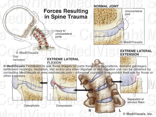
A traumatic event causing injury to an intervertebral disc may also cause subtle injuries to the bones around the disc. During an extreme lateral flexion injury (shown in the image below), the edges of the bone are driven together, injuring both the disc and the bone. As the bone/disc junction heals, overgrowths referred to as osteophytes may form.

Osteophyte Formation From Trauma

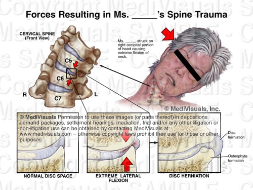
Osteophytes take weeks or months to develop following a traumatic event; therefore, any osteophytes that are present soon after a traumatic event are likely pre-existing.

Cervical Spine Osteophytes

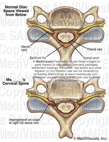
The osteophytes themselves may compress the neural elements as in the illustration above; however, in most situations, the osteophytes are a part of an OSTEOPHYTE/DISC COMPLEX. This is when the osteophytes and disc extend beyond their normal limits and compress the neural elements (spinal cord, nerve roots). In cases where osteophytes may have pre-existed a traumatic event, worsening of the disc bulge could occur following the trauma, resulting in new or aggravated symptoms.

Sagittal Spine Osteophyte Disc

Sometimes disc and ligament injuries occur on the same side as the force of impact. Other times, they occur on the opposite side. Injuries to the disc on the same side as the force are the result of stretching and tearing forces. On the opposite side, compression forces result in tears and micro fractures of the tissues and bones. (see illustration below). Osteophytes and facet hypertrophy can also form following injuries to intervertebral discs and ligaments. Injuries to the discs and ligaments result in instability and excessive motion of the joints that, in turn, results in constant trauma to the bone/disc and ligament junctions. This ongoing trauma results in overgrowth of the bones as it continually cycles through episodes of healing and reinjury.

Spine Trauma Forces Osteophyte Disc

Topics: mechanism of injury, intervertebral disc, hyperflexion, trial exhibit, trauma, disc injury, personal injury, osteophyte

Understanding the Brachial Plexus Injury: Part 2 (Shoulder Dystocia)

Posted by Trisha Haszel Kreibich on Fri, Nov 5, 2010
By: Trisha Kreibich, MS, Medical Illustrator/Consultant, and Robert Shepherd, MS, CMI, Medical Illustrator, Executive Vice President and Operations Director, Eastern Region MediVisuals Inc

This article is a continuation of a two part article on brachial plexus injury. Part 1 covered brachial plexus injuries in adults caused by a traumatic event, such as a motor vehicle collision. Part 2 will address brachial plexus injury in infants during delivery, which is also known as Shoulder Dystocia or Erb's palsy.

To review, the brachial plexus innervates the arm and is formed by several of the cervical nerve roots and the T1 nerve root. [see illustration below]

Pediatric Brachial Plexus

During delivery, the infant's passage through the birth canal can be stopped by the impaction of its shoulder(s) against the mother's pubic bone, sacral promontory, or both. [see illustration below]

Shoulder Dystocia

Injury to the brachial plexus may sometimes occur because of unusually powerful uterine contractions or rapid fetal descent; however, brachial plexus injury may also result from improper obstetrician interaction. When excessive downward traction on the head of an infant with shoulder dystocia is applied (a violation of the standard of care), the delicate brachial plexus is stretched and injured. [shown below] The same may also occur when vacuum extraction is used in this situation.

Shoulder Dystocia and Excessive Traction

As in adult brachial plexus injuries, the earlier the injury is identified and treated, the better chances are for recovery and preventing permanent damage. However, this is also dependent on the severity of the initial injury. Sometimes, an overgrowth of nerve cells (neuroma) can form around the injured nerves. When this occurs, surgical intervention and nerve grafting may be needed. [see illustration below]

Sural Nerve Graft

There are some delivery techniques that can be used to prevent brachial plexus injury in infants with shoulder dystocia. As the illustration below demonstrates, the mother is moved to the McRoberts position, where her legs are hyperflexed to the abdomen, typically resulting in an increase of the outlet. This is often used in conjuction with an episiotomy, a cut made in the perineal body (tissue between the vagina and anus) before delivery, to enlarge the outlet and allow the obstetrician more room to perform maneuvers.

McRoberts Position

If no progress is made, moderate suprapubic pressure may be applied to free the impacted shoulder. The Wood's screw maneuver [shown below] may be used as well. This maneuver involves the obstetrician rotating the infant's anterior or posterior shoulder, and in turn the body, like a screw, freeing the impacted shoulder.

Woods Screw Maneuver 604058 03X

The following animation was developed to show how brachial plexus injuries can occur in a shoulder dystocia case, along with a few of the above mentioned accepted procedures that can be performed to help prevent this injury.

Shoulder Dystocia Animation

Reference:

Gabbe, S.G., Niebyl, J. R., & Simpson, J.L. "Obstetrics: Normal & Problem Pregnancies." 3rd ed. Philadelphia: Churchill Livingstone, 1996. 374-375, 490-494. Print.

Topics: mechanism of injury, brachial plexus, Erb's palsy, trial exhibit, medical-legal-illustration, birth trauma, medical exhibit

Vestibular Nerve Injury: Why it could be important to your TBI case

Posted by Trisha Haszel Kreibich on Tue, Oct 5, 2010

By: Robert Shepherd MS, Certified Medical Illustrator, Vice President and Director of Eastern Region Operations, MediVisuals Incorporated

Balance and dizziness are often associated with traumatic brain injuries, although the specific cause of these problems is often difficult to explain. Sometimes the injuries may be to the inner ear organs. Other times the injuries may be to the vestibular nerve. When the injury is to the vestibular nerve, the mechanism of injury is similar to injuries to the olfactory nerve resulting in disturbances in smell.

The exhibit shown below demonstrates the mechanism of injury. As the brain stem and skull move in different directions during a violent impact, stretch injuries to the vestibular nerve can occur. This type of injury is especially significant when supporting arguments of brain injuries occurring as a result of traumatic forces to the head. If forces were significant enough to damage the vestibular nerve, the forces were likely sufficient to cause shear or traumatic axonal injury, as well.

Vestibular Nerve Injury 500

For more information on mild and severe traumatic brain injury, please visit: http://www.medivisuals.com/traumatic-brain-injury.aspx For more information on the featured exhibit, please visit: http://www.medivisuals.com/vestibular-nerve-injury-mvi82010-01x.aspx

Topics: mechanism of injury, trial exhibit, traumatic-brain-injury, medical-legal-illustration, vestibular nerve

Soft Tissue Injury - Cervical and Lumbar Strain

Posted by Delia Dykes on Fri, Mar 12, 2010

By: Robert Shepherd MS, Certified Medical Illustrator, Vice President and Director of Eastern Region Operations, MediVisuals Incorporated 

Injuries to the spinal and paraspinal ligaments and muscles can result from violent side-to-side motions or by violent excessive flexion and extension. The illustration below shows the major ligaments of the neck (anterior longitudinal and interspinal ligaments) in hyperflexion and hyperextension, which can be injured grossly or microscopically.

cervical-strain-injury

Click to enlarge

The series of three illustrations in the bottom right corner (which is shown in more detail below), show a close-up view of the spinal anatomy in 1.) the Normal condition, 2.) during Excessive Stretching and 3.) After Healing. In the Normal condition, one can appreciate the close relationship between the muscles, nerves and blood vessels. During Excessive Stretch, small tears occur, which causes bleeding in the muscle fibers. After Healing, scar tissue and inflammation entrap blood vessels and nerves resulting in a permanent state of compromised, painful movement.

muscle-stretched
 

The same is true for the lower lumbosacral spine and pelvic regions. In the illustration below, the spinal nerves and their posterior branches are seen in close approximation to the ligaments and joint capsules, which are often involved in the injury. During hyperflexion of the lumbar spine, transient bulging of the intervertebral discs can occur. 


lumbar-strain-injury

Click to enlarge

The series of illustrations in the lower right corner of the above image, show the normal lumbosacral and pelvic muscles and tendon fibers, which insert on the bones near the associated posterior spinal nerve branches. During Excessive Stretch, a segmental artery and its branches may be involved in hemorrhaging, scarring and occlusion. After Healing, scar tissue and adhesions form, entrapping nerves and blood vessels, causing chronic pain.

In these next illustrations, muscle is shown in sequentially higher magnifications, which can be used to explain excessive stretch injuries in any area of the body.  In the Normal series, the bottom illustration depicts nerves and small body vessels intertwined in muscle with its tendinous attachment to the bone.  The middle illustration shows a magnified view of an individual muscle fiber and the top illustration depicts the relationship of the microscopic myofilaments (actin and myosin) in their normal relaxed position.

 muscle-normal


In the Excessive Stretch series, the bottom illustration shows the muscle, tendon, blood vessels and nerves as they are excessively stretched. Small hemorrhages are seen escaping from the stretched and torn blood vessels. In the middle illustration, blood is shown escaping into surrounding spaces, reducing oxygen exchange to the muscle and irritating the delicate structures of the muscle fiber. The top illustration depicts the myofilaments, showing the actin and myosin fibers torn and stretched past the point of normal interdigitation.

 muscle-excessive-stretch


In the After Healing series, the bottom illustration shows the irregular outline of the scarred and inflammed muscle fiber with small adhesions seen between the blood vessels, nerves and muscle fibers. The middle illustration shows scar tissue and inflammation occluding blood vessels and adhering the delicate structures of the muscle fibers together, limiting motion and causing chronic pain. Lastly, the top illustration depicts the damaged myofilaments. Their normally well-organized, interdigitating arrangement is left destroyed, limiting muscle movement at the most basic level.

muscle-after-healing
   

Topics: mechanism of injury, coup-contracoup, demonstrative evidence, soft tissue injury, hyperflexion, lumbar strain, hyperextension, medical-legal-illustration, medical expert, spinal injury, cervical strain, connective tissue