Medical Exhibits - Demonstrative Evidence Expert Blog - MediVisuals

Medical-Legal Illustrators Judged Best in Profession

Posted by Trisha Haszel Kreibich on Thu, Aug 19, 2010

There may be no better measuring stick for an attorney than the opinions of their peers. The same is true for medical illustrators.

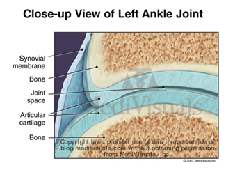
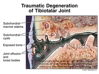
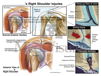
During the Association of Medical Illustrators (AMI) Annual Conference, each year a judged illustration competition is held. One of the largest and most competitive categories is the Medical-Legal category. Unlike most art competitions, the illustrations are not judged primarily on aesthetics. Instead, the most important criteria are anatomical accuracy and how effective the visuals are in communicating a stated message to the jury.

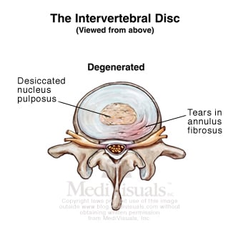
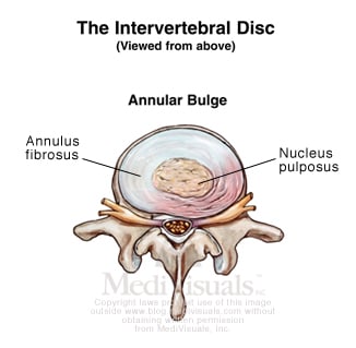
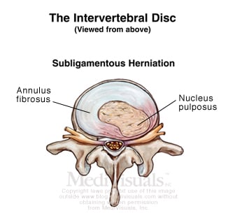
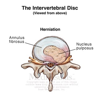
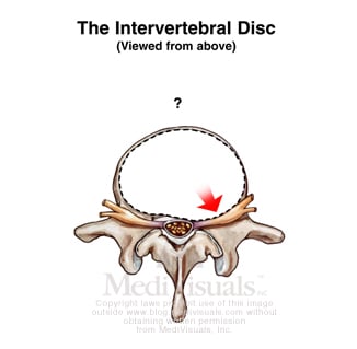
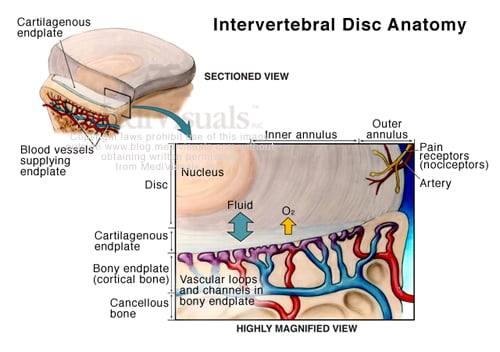
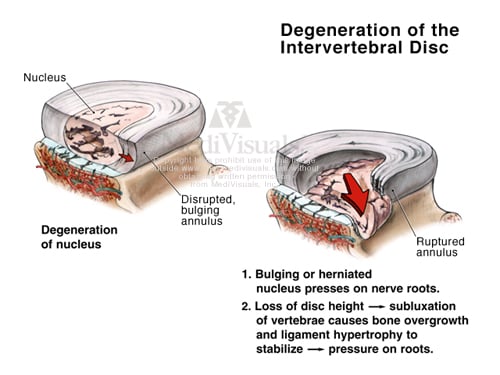
At the recent 2010 Conference in Portland, Oregon, the illustrations in the medical-legal category were particularly strong, resulting in the presentation of two Awards of Excellence (the highest honor) and four Awards of Merit. Illustrators with MediVisuals of Richmond, Virginia and Dallas, Texas were honored with both the Awards of Excellence and three of the four Awards of Merit. The various award winning illustrations can be seen by clicking the following link: http://www.medivisuals.com/awards.aspx.

The Award of Excellence in Medical Legal Illustration is particularly significant because this Award has only been bestowed 23 times in the 75 year history of the AMI. With this year’s Awards, MediVisuals has now received 18 of the 23.

MediVisuals was also honored with an Award for a medical-legal animation entitled "Cascade Effect of Axonal Loss". The animation was developed for use in all "mild" TBI cases and helps explain how injury to even one axon can interfere with the ability of several neurons to communicate with each other. The animation can be viewed using the following link or clicking the image below: Cascade Effect of Single Axon Loss Animation

For more information on MediVisuals or to receive a complimentary consultation on an upcoming case, please visit www.medivisuals.com

Cascade Animation Screen Blog


By: Robert Shepherd, MS, Certified Medical Illustrator, Vice President and Director of Eastern Region Operations, MediVisuals Incorporated

Topics: medical-illustrator, medical-legal-illustration, MediVisuals

Soft Tissue Injury - Cervical and Lumbar Strain

Posted by Delia Dykes on Fri, Mar 12, 2010

By: Robert Shepherd MS, Certified Medical Illustrator, Vice President and Director of Eastern Region Operations, MediVisuals Incorporated 

Injuries to the spinal and paraspinal ligaments and muscles can result from violent side-to-side motions or by violent excessive flexion and extension. The illustration below shows the major ligaments of the neck (anterior longitudinal and interspinal ligaments) in hyperflexion and hyperextension, which can be injured grossly or microscopically.

cervical-strain-injury

Click to enlarge

The series of three illustrations in the bottom right corner (which is shown in more detail below), show a close-up view of the spinal anatomy in 1.) the Normal condition, 2.) during Excessive Stretching and 3.) After Healing. In the Normal condition, one can appreciate the close relationship between the muscles, nerves and blood vessels. During Excessive Stretch, small tears occur, which causes bleeding in the muscle fibers. After Healing, scar tissue and inflammation entrap blood vessels and nerves resulting in a permanent state of compromised, painful movement.

muscle-stretched
 

The same is true for the lower lumbosacral spine and pelvic regions. In the illustration below, the spinal nerves and their posterior branches are seen in close approximation to the ligaments and joint capsules, which are often involved in the injury. During hyperflexion of the lumbar spine, transient bulging of the intervertebral discs can occur. 


lumbar-strain-injury

Click to enlarge

The series of illustrations in the lower right corner of the above image, show the normal lumbosacral and pelvic muscles and tendon fibers, which insert on the bones near the associated posterior spinal nerve branches. During Excessive Stretch, a segmental artery and its branches may be involved in hemorrhaging, scarring and occlusion. After Healing, scar tissue and adhesions form, entrapping nerves and blood vessels, causing chronic pain.

In these next illustrations, muscle is shown in sequentially higher magnifications, which can be used to explain excessive stretch injuries in any area of the body.  In the Normal series, the bottom illustration depicts nerves and small body vessels intertwined in muscle with its tendinous attachment to the bone.  The middle illustration shows a magnified view of an individual muscle fiber and the top illustration depicts the relationship of the microscopic myofilaments (actin and myosin) in their normal relaxed position.

 muscle-normal


In the Excessive Stretch series, the bottom illustration shows the muscle, tendon, blood vessels and nerves as they are excessively stretched. Small hemorrhages are seen escaping from the stretched and torn blood vessels. In the middle illustration, blood is shown escaping into surrounding spaces, reducing oxygen exchange to the muscle and irritating the delicate structures of the muscle fiber. The top illustration depicts the myofilaments, showing the actin and myosin fibers torn and stretched past the point of normal interdigitation.

 muscle-excessive-stretch


In the After Healing series, the bottom illustration shows the irregular outline of the scarred and inflammed muscle fiber with small adhesions seen between the blood vessels, nerves and muscle fibers. The middle illustration shows scar tissue and inflammation occluding blood vessels and adhering the delicate structures of the muscle fibers together, limiting motion and causing chronic pain. Lastly, the top illustration depicts the damaged myofilaments. Their normally well-organized, interdigitating arrangement is left destroyed, limiting muscle movement at the most basic level.

muscle-after-healing
   

Topics: mechanism of injury, coup-contracoup, demonstrative evidence, soft tissue injury, hyperflexion, lumbar strain, hyperextension, medical-legal-illustration, medical expert, spinal injury, cervical strain, connective tissue

Disc Herniation and Other Disc Injuries

Posted by Trisha Haszel Kreibich on Wed, Jan 20, 2010

By: Robert Shepherd MS, Certified Medical Illustrator, Vice President and Director of Eastern Region Operations, MediVisuals Incorporated 

The term “degenerated disc” is generally used to describe a disc in the early degenerative process.  It is the beginning of a progressive break down of the disc. This condition can be initiated or accelerated by a traumatic event.

degenerated-disc

A disc bulge is a more advanced collapse of the disc to the point that the disc expands beyond its normal contour. It may or may not impinge on the neural structures within the spinal canal or neural foramina. Similarly, a disc bulge can be the immediate or delayed result of a traumatic event, or a traumatic event may exacerbate a preexisting, stable disc bulge.

annular-disc-bulge

A subligamentous herniation is one in which nucleus pulposus has extended through the annulus fibrosus, but has not gone through the posterior longitudinal ligament.

subligamentous-herniation

The term “herniation” is generally used when the nucleus has completely extruded through the annulus fibrosus and posterior longitudinal ligament.

However, it does not matter what the disc pathology is labeled, if it impinges upon, or irritates the neural components, it is a significant injury that will likely require some type of invasive procedure to correct.

 impinge-disc-injury

The disc can either be injured by an immediate tear of the annulus fibrosus and extrusion of the nucleus pulposus during a traumatic event, or they can be the result of a much more gradual process. In order to understand the gradual breakdown of the disc, one must first understand a little of the physiology of a disc. The inner disc relies on exchange of fluid, nutrients and oxygen through the end plate of the adjacent vertebral bodies.

disc-anatomy

During a traumatic event, the endplate may become injured resulting in interference with that exchange.

disc-disruption

As a result of the inability of the disc to obtain the fluid, nutrients, and oxygen it needs, the disc gradually begins to break down - becoming a degenerated disc, followed by a bulging disc, and eventually to a herniated disc. The amount of time involved with the process of the disc breakdown is related to the severity of the initial disc injury.

degeneration-of-disc

Therefore, if a disc injury is not evident until weeks or months after a traumatic event, it does not mean the injury was not a direct result of the traumatic event.


Topics: disc herniation, degenerated disc, medical-legal-illustration, MediVisuals

Breaking Down Traumatic Arthritis

Posted by Trisha Haszel Kreibich on Tue, Dec 15, 2009

By: Robert Shepherd MS, Certified Medical Illustrator, Vice President and Director of Eastern Region Operations, MediVisuals Incorporated

Fractures can result in several long term or permanent complications that can necessitate additional surgical procedures. One of the most common long term debilitating complications is traumatic arthritis. Traumatic arthritis can affect almost any moveable joint in the body. To explain traumatic arthritis more in depth, we will be focusing on the tibiotalar (ankle) joint, as shown in the illustration to the right.

arthritis-tibiotalar-joint

As like most moveable joints, the tibiotalar joint consists of smooth articular bone covered by thick, shock-absorbing articular cartilage.

arthritis-joint-normal

During trauma, the joint surfaces can be driven together resulting in injuries to the cartilage and microfractures of the articular surface (even without obvious intrarticular fracture).

arthritis-injury-joint

The bone and cartilage then undergo changes that result in the progressive breakdown of the joint. As the process advances, the joint becomes painful. In most cases, the only treatment options are joint replacement or fusion.

arthritis-degeneration-joint

The acromioclavicular (AC) joint also frequently falls victim to traumatic arthritis. As the AC joint enlarges (hypertrophy) it impinges on the rotator cuff, which is referred to as subacromial impingement. This can cause irritation or tearing of the rotator cuff and is most often treated by AC joint resection and subacromial decompression.

arthritis-AC-joint-shoulder


ZSN7H32E27UR

Topics: arthritis, joint, ankle, medical-legal-illustration, MediVisuals

Selecting a Medical Illustrator

Posted by Trisha Haszel Kreibich on Fri, Sep 25, 2009

By: Robert Shepherd MS, Certified Medical Illustrator, Vice President and Director of Eastern Region Operations, MediVisuals Incorporated

Just as all treating physicians and/or medical experts are not the same, all medical illustrators are not the same. Attorneys typically go to great lengths to ensure that testifying experts for their cases are adequately credentialed, but many give little thought to applying the same due diligence when selecting a medical illustrator. This can be a big mistake because unlike many “professionals”, there is no basic training, licensing, or certification process that is required for an individual to call themselves a “medical illustrator”. Many less skilled and/or less qualified “medical illustrators” market to attorneys – perhaps because attorneys are less able to detect errors in their work. For these reasons, it is the responsibility of the attorney and/or medical expert to their clients to ensure they enlist the services of a medical illustrator who is qualified to provide those services.

Working with a qualified medical illustrator who has experience in the medical-legal area can be a pleasant, enjoyable experience for the testifying expert as these medical illustrators are able to read and comprehend medical records, review imaging studies, and discuss complicated anatomical and medical terms on a very similar level as the expert. On the other hand, working with an un- or minimally qualified and/or inexperienced medical illustrator can be a frustrating and time consuming task that may require multiple revisions of drafts, ineffective demonstrative aids, or even embarrassment during a hearing because of the discovery of some error or inconsistency during testimony.

It can be difficult to determine the qualifications of a medical illustrator based upon looking at their artwork alone. A copy of a résumé or curriculum vitae should be requested. One of the most basic requirements that the résumé should show would include graduation from one of the below medical illustration graduate programs. There are several medical illustration programs in colleges and various institutes in North America and across the globe; however, only five are currently accredited by the American Medical Association, and Commission on Accreditation of Allied Health Education Programs. Those programs include:

  • Medical College of Georgia - Master of Science in Medical Illustration

  • University of Illinois at Chicago - Master of Science in Biomedical Visualization

  • Johns Hopkins University - Master of Arts in Medical and Biological Illustration

  • University of Texas, Southwestern – Master of Arts in Biomedical Communications

  • University of Toronto - Master of Science in Biomedical Communications

Other criteria that should be evident in a résumé or CV that would help demonstrate at least minimal qualifications are 1) Certification as a medical illustrator as issued by The Board of Certification of Medical Illustrators and 2) Professional Membership in the Association of Medical Illustrators. Both of these require that the individual possess at least minimal medical illustration training and skill levels. Also, just as there are different subspecialties in medicine which require specific and advanced skills and knowledge, specialization in “medical-legal” illustration requires additional knowledge and experience in addition to those required for general medical illustration. For that reason, it is also wise to select a qualified medical illustrator who is not only familiar with illustration and medicine, but also has a significant amount of experience in creating illustrations for litigation purposes.


Topics: medical-illustrator, medical-legal-illustration, MediVisuals

Medical Illustrators as a Member of the Medical-Legal Team

Posted by Trisha Haszel Kreibich on Thu, Sep 24, 2009

By: Robert L. Shepherd MS, Certified Medical Illustrator, Vice President and Director of Eastern Region Operations, MediVisuals Incorporated

A very effective way of increasing the effectiveness of expert testimony is by enhancing focus, understanding, and recall by teaming the testifying expert with a qualified medical illustrator, experienced in preparing legal graphics (illustrations, animations, models, etc.). By working together, the medical knowledge and oral skills of the expert can be supported by expertly created medical graphics that greatly clarify complicated anatomical, physiological, and medical subtleties. Together they result in much more effective communication with the decision makers than if verbal testimony or the graphics were used alone (one mode of communication used alone without being supported by the other). In addition, during a period of hours or days of listening to arguments that are typically only verbal, decision makers grasp the opportunity to focus on visuals in the form of illustrations, photographs, models, and/or animations. In fact, information is generally better processed if jury members and other triers of fact can have information presented in a multimodal fashion (i.e. combinations of simultaneous auditory, visual, and tactile stimuli).

The effectiveness of visuals is supported throughout our society by such common phrases as “A picture is worth a thousand words”, and “Seeing is believing.” In addition, numerous manuscripts refer to studies substantiating that recall is greatly increased when the verbal message is supported by visual images. Typically these studies have shown that after varying periods of time information that was delivered by a combination of voice supported by visuals was recalled at a significantly higher percentage than the same message delivered by voice alone.

References

DeBoth, C. J., & Dominowski, R. L. Individual differences in learning: Visual versus
auditory presentation. Journal of Educational Psychology, 1978; Aug 70 (4): 498-503

McCall, J., & Rae, G. Relative efficiency of visual, auditory and combined modes of
presentation in learning of paired-associates. Perceptual and Motor Skills, 1974; June (38) : 955-958.

Multimodal Learning Through Media: What the Research Says. Cisco Web site. http://www.cisco.com/web/strategy/docs/education/Multimodal-Learning-Through-Media.pdf. Accessed August 19, 2008.


Topics: medical-illustrator, traumatic-brain-injury, medical-legal-illustration, MediVisuals